A FEW NOTES ON BIONEUROPHYSIOLOGY
| Books - The Knowledge of the Womb |
Drug Abuse
CHAPTER II
A FEW NOTES ON BIONEUROPHYSIOLOGY
§ 86 The nervous system consists of a large number of nerve cells (neurons) which are separated from each other by synapses.
The direct result of the excitation of a living neuron 'n' by a stimulus 's' is the secretion of neurotransmittter into the synapses which separate the axon terminals of 'n' from the dendrites and/or cell body of 'n's postsynaptic neurons. (In this case, 'n' is
a presynaptic neuron.3) Whether Vs excitation by 's' is transmitted to some or all of 'n's postsynaptic neurons or whether it is not transmitted to any postsynaptic neuron, depends on the quality and intensity of V.
As far as the quality of a stimulus is concerned, Eric Kandel's experiments on the Aplysia Californica 4 show that this mollusc withdraws from a harmful stimulus thanks to the transmission of the excitation of presynaptic neurons (in this case, sensory) to their postsynaptic neurons (in this case, motor). On the other hand, the mollusc hardly reacts to a stimulus which is neither harmful nor beneficial because the excitation of presynaptic neurons (again, sensory) is not transmitted to postsynaptic neurons (again, motor). Kandel's experiments show that even such a simple organism as the Aplysia Californica has the ability to recognize the biological significance of stimuli which excite it.
With what mechanism do the sensory neurons of the Aplysia Californica recognize the biological significance of stimuli which excite it?
§ 87 The role played by the intensity of a stimulus in the transmission of neuronal excitation may be seen in the following example. When the syllable o and vo are uttered in an audible voice, they constitute acoustic stimuli. If these acoustic stimuli are repeated frequently at equal intervals and with the same intensity, they will combine in the following way: ...μo-vo-μo-vo-μo-vo-μo-vo-μo-vo-μo-vo-μo-vo-μo-vo-μo ... As a result, the nervous system will be unable to distinguish which of the following four words excited it: μό-vo,μo-vό, vό-μo or vo-μό5 The distinction will depend on there being a pause following a single combination of the stimuli μo and vo, on the chronological order of the combined stimuli and on the intensity of the stimuli.
If the stimulusμo is followed by the stimulus vo and then there is a pause, the combination will give the wordμo-vo, but which of the two words will it be, μό-vo or μo-vό? For the nervous system to distinguish between these two words, one of the two stimuli must be stressed, that is, it must be of greater intensity.
What is the result when the intensity of a stimulus exciting a neuron increases? The answer is given by H. K. Hartline's experiemental findings6 (fig. 1). These show that when the instesity of a stimulus increases, the number of impulses which a neuron presents in a unit of time also increases. In my view, this process in turn increases the quantity of a neurotransmitter secreted into the synapses of the excited neuron and results in the transmission of the excitation to a greater number of postsynaptic neurons. The threshold of different qualities of neurons varies (§ 88): the higher the threshold of a neuron, the larger the quantity of neurotransmitter needed to excite it. Thus, the greater the intensity of a stimulus exciting a presynaptic neuron, the greater the number and the more qualities of postsynaptic neurons which are excited (see figures 2, 3, 4, 5, and 6).
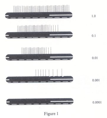
Figure 1 Impulses set up in optic nerve fibre of Limulus by one second flash of with relative intensities shown at right. The lower white line marks 0.2 second v vats and the gap in the upper white line gives the period for which the eye was ill nated. (From Hartline, 1934.)
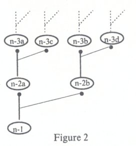
Figure 2 For simplicity's sake, a peripheral sensory neuron, n-1, is presented as linking up with only two neurons, n-2a and n-2b, each of different quality to the other. Each one of the latter (again for simplicity) links up with only two neurons - n-3a and n-3c; n-3b and n-3d - each of different quality to the other.
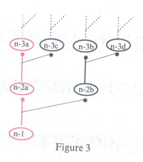
Figure 3 shows the specific neuronal path or circuit along which the excitation from neuron n-1 is transmitted when n-1 has been excited by a stimulus's' whose intensity is one degree. The transmission of the excitation from n-1 to n-2a and then to n-3a produces a specific result. The neurons which are excited are marked in red.
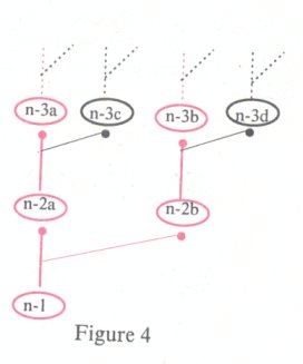
Figure 4 shows the specific path-circuit along which the excitation from neuron n-1 is transmitted when n-1 has been excited by the same stimulus 's' (ie. its quality remains constant) whose intensity is now double, that is, two degrees. The transmission of the excitation from n-1 to n-2a, n-2b, n-3a and n-3b produces a specific result which is different to that of figure 3. The neurons which are excited are marked in red.
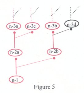
Figure 5 shows the specific path-circuit along which the excitation from neuron n- I is transmitted when n-1 has been excited by the same stimulus's' whose intensity has now tripled, that is, to three degrees. The transmission of the excitation to n-2a, n-2b, n-3a, n-3c and n-3b produces a specific result which is different to that of figures 3 and 4. The neurons which are excited are marked in red.
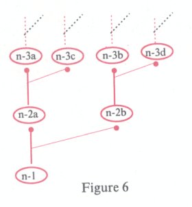
Figure 6 shows the specific path-circuit along which the excitation from neuron n- I is transmitted when n-1 has been excited by the same stimulus's' whose intensity has now quadrupled, that is, to four degrees. The transmission of the excitation to n-2a, n-2b, n-3a n-3c, n-3b and n-3d produces a specific result which is different to that of figures 3, 4 and 5. The neurons which are excited are marked in red.
If figures 3 - 6 we can see that when neuron n-1 is excited by a stimulus constant in quality but different in intensity, the excitation follows four different and specific neuronal paths or circuits through the nervous system. Of course, the specific result of each path-circuit will be different, depending on the quality of the neurons which are excited at the various levels of the nervous system: the spinal cord, cerebral stem, diencephalon, limbic system and the cortex of the cerebral hemispheres (see § 89 for more details).
The result of combinations of stimuli, such as μo and vo, will also depend, as we have said, on their chronological order.
In figure 7, for simplicity's sake:
(a) Neuron μo- 1 symbolizes all the specific peripheral acoustic neurons which are excited by the acoustic stimulus po.
(b) Neuron vo-1 symbolizes all the specific peripheral acoustic neurons which are excited by the acoustic stimulus vo.
(c) Neurons μo-1,μo-2a, μo-2b, vo-1, vo-2a and vo-2b are each presented as joining up with only two postsynaptic neurons. (In reality every neuron links up with a multitude of postsynaptic neurons of different quality).
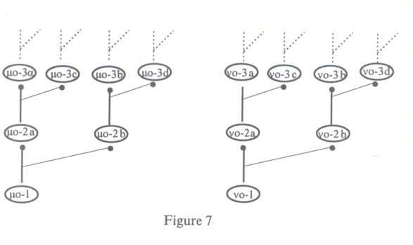
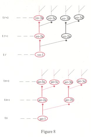
Figure 8 This shows the specific neuronal path or circuit along which the excitation from neuronsμo-1 and vo-1 is transmitted whenμo-1 and vo-1 have been excited by the acoustic stimuli μόand vo, in that order. The result is the acoustic impressionμόvo. The neurons which are excited are marked in red. Because the stimulusμό precedes the stimulus vo, it excites neuron μo-1 before the stimulus vo excites vo-1, as shown in the figure ('t' stands for time). Because the stimulus μο is stressed, its intensity is greater than that of vo: thus the excitation fromμo-1 is transmitted to neuronsμo-2a, μo-2b,μo-3a, μo-3c,μo-3b and μo-3d. Because the stimulus vo is unstressed, its intensity is less than that ofμo and thus the excitation from vo-1 is transmitted only to neurons vo-2a and vo-3a.
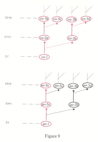
Figure 9 This shows the specific path-circuit along which the excitation from neuronsμo-1 and vo-1 is transmitted when these neurons have been excited by the acoustic stimuli μo and vό, in that order. The result is the acoustic impression μovό. The neurons which are excited are marked in red. Because the acoustic stimulusμo precedes the acoustic stimulus vό, it excites neuron μo-1 before the stimulus vό excites vo-1, as shown in the figure. Because the stimulusμo is unstressed, its intensity is less than that of vό and thus the excitation fromμo-1 is transmitted only to neuronsμo-2a and μo3a. Because the stimulus vό is stressed, its intensity is greater than that of μo: thus the excitation from vo-1 is transmitted to vo-2a, vo-2b, vo-3a, vo-3c, vo-3b and vo-3d.
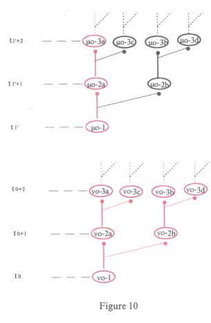
Figure 10 This shows the specific path-circuit along which the excitation from neurons vo-1 andμo-1 is transmitted when these neurons have been excited by the acoustic stimuli vό andμo, in that order. The result is the acoustic impression vόμo. The neurons which are excited are marked in red. Because the acoustic stimulus vό precedes the acoustic stimulus μo, it excites neuron vo-1 before the stimulus excites μo-1, as shown in the figure. Because the stimulus vό is stressed, its intensity is greater than that of μo: thus the excitation from vo-1 is transmitted to neurons vo-2a, vo-2b, vo3a, vo-3c, vo-3b and vo-3d. Because of the stimulusμo is unstressed, its intensity is less than that of vό and thus the excitation fromμo-1 is transmitted only to neuronsμo-2a and μo-3a.
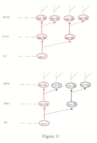
Figure 11 This shows the specific path-circuit along which the excitation from neurons vo-1 andμo-1 is transmitted when these neurons have been excited by the acoustic stimuli vo and μό, in that order. The result is the acoustic impression voμό. The neurons which are excited are marked in red. Because the acoustic stimulus vo precedes the acoustic stimulus μό, it excites the neuron vo-1 before the stimulus μo excites μo-1, as shown in the figure. Because the stimulus vo is unstressed, its intensity is less than that ofμό and thus the excitation from vo-1 is transmitted only to neurons vo-2a and vo-3a. Because the stimulus μό is stressed, its intensity is greater than that of vo: thus the excitation fromμo-1 is transmitted to neuronsμo-2a, μo-2b,μo-3a, μo-3c,μo3b and μo-3d.
In figures 8 - 11, we can see that the excitation of neurons μo-1 and vo-1 by the acoustic stimuli μo and vo follows four different neuronal path-circuits and produces four different results or acoustic impressions according to the intensity of the stimuli and the chronological order in which they excite μo-1 and vo-l.
Note: Of basic importance to the functioning of the nervous system is the ability of a neuron to participate in many neuronal circuits, figures 8, 9, 10 and 11, neuronμo-2a participates in four different circuits with a different result in each case.
§ 88 A most basic factor which influences the quality and intensity of a nervous system's reaction at a given moment is the threshold of its neurons at that same moment. One factor which regulates the threshold of neurons is their metabolism. For example, a dog which has just satiated its hunger does not present the same degree of salivation at the sight of food as it would if it were hungry. (This observation shows that experimental research into the process of neuronal excitation should be carried out concurrently with research into the process of metabolism occurring at the same moment as the excitation.)
The threshold of neurons also depends on their quality. For example, pyramidal motor neurons have a different threshold to that of spinal motor neurons.
§ 89 The role played by the quality of the neurons which participate in a pathcircuit of neuronal excitation can be seen in the following:
(a) Through sensorial neurons the excitation results in sensorial symptoms of sight, hearing, touch, kinaesthesia and so on.
(b) Through motor neurons (pyramidal cells and motor neurons of the anterior horns of the spinal cord) the excitation results in the contraction of striated muscles.
(c) Through neurovegetative neurons it results in neurovegetative S & P.
(d) Through neurons of the limbic system it results in emotional symptoms of fear, anger and so forth.
(e) Through neurons of the frontal poles it results in the symptom of streams of thought.
(f) Through the existential neurons the excitation results in the symptom of consciousness of existential identity.
Note: The clinical data of Petit Mal fits, postconcussional and postepileptic automatism, sleep, fainting attacks and other states support the view that R's nervous system contains a special and highly complex neuronal circuit whose excitaion gives rise to the symptom of consciousness of existential identity. For the sake of brevity, the special circuit is called 'existential neurons'. When R's existential neurons are inhibited, so too is the symptom of consciousness of existential identity: in other words, R ceases to exist for himself. (Dreams support the viewpoint that the existential neurons may be excited during sleep.)
§ 90 Recapitulation of § 86 - 89: The excitation of a neuron 'n' by a stimulus 's' at a given moment is transmitted successively to a series of neurons of the same and different quality. The end result of this entire process depends on:
(a) The quality and intensity of V. (b) The quality of V.
(c) The threshold - at the same moment - of the neurons which connect directly or indirectly with 'n'.
(d) The quality of the series of neurons to which the excitation of 'n' is transmitted.
(e) When many neurons are excited by different stimuli the end result also depends on the chronological order of their excitation (figs. 8 - 11).
3 The terms 'presynaptic' and 'postsynaptic neurons' refer to the relationship between any neurons which link up with each other. Any neuron can be presynaptic. Postsynaptic neurons are those neurons whose dendrites and/or cell body link up with the axon terminals of one or more presynaptic neurons. In fig 2 (p.175), for example, neuron n- I is presynaptic to neurons n-2a and n-2b which are postsynaptic to n-1: neuron n-2a is presynaptic to neurons n-3a and n-3c which are postsynaptic to n-2a, while n-2b is presynaptic to n-3b and n-3d which are postsynaptic to n-2b, and so on.
4 Eric Kandel, "Small Systems of Neurons", Scientific American, 'The Brain', Sept. 1979.
5 Translator's note: The Greek wordsμόvo,μοvό, vόμo and voμόhave been retained in the English text.μόvo, with the stress on the first syllable as shown by the accent, means 'alone'.μοvό, with the stress on the second syllable as shown by the accent, means 'odd' as in 'odd and even numbers', or 'single' as opposed to 'double'. vόμο with the stress on the first syllable means 'law' while voμό with the stress on the second syllable means 'prefecture' or 'county'.
Each of these words has a peculiarity that does not exist in English, namely that by interchanging the consonants (μ and v) of the first and second syllables (μo and vo) as well as the emphasis (from the first to the second syllable and vice versa) - the vowel of each syllable being a constant 'o' - three more words are produced.
6 H. K. Hartline, "Intensity and Duration in the Excitation of Single Photoreceptor Units", J. Cell. Comp. Physiol. 5, 229, 1934.
| < Prev | Next > |
|---|


